




|
Quantity
|
Out of stock
|
||
|
|
|||
|
|
|||
Animal and Internal Organ Models: Essential Tools for Veterinary Training
Understanding the internal anatomy of animals is crucial in veterinary education and clinical practice. Our veterinary animal and organ models offer a detailed, hands-on approach to learning, helping students, veterinarians, and educators visualise the structure, position, and function of internal systems. Veteq Group provides a range of professional-grade anatomical models for use in vet schools, clinics, and training centres across the UK.

Why Choose Animal and Organ Models for Education?
These models support learning across multiple settings—from classrooms and anatomy labs to clinics and client consultations. They allow veterinary students to study anatomical structures without the need for preserved specimens, while veterinarians can use them to explain complex conditions to pet owners more clearly. Our range ensures high anatomical accuracy and ethical, reusable alternatives to traditional methods.
Key Benefits of Anatomical Models
- Durability: Made from high-strength plastics for long-term use, even in demanding teaching environments.
- Realistic appearance: Accurate colours, textures, and anatomical proportions replicate real animal tissues and organs.
- Low maintenance: No refrigeration or preservatives needed. Easy to clean and store.
- Safe and hygienic: Made from non-toxic, skin-safe materials that pose no biological hazard.
- Affordable: Competitive pricing makes these models accessible for clinics, schools, and organisations of all sizes.
- Space-saving: Mounted on solid wooden bases for secure display and easy shelf storage.
- Lightweight and ergonomic: Easy to handle, inspect, and demonstrate from any angle.
Our Veterinary Model Selection Includes:
- Full animal models: Including cats, rabbits, dogs, goats, and horses—ideal for studying external anatomy with optional internal cross-sections.
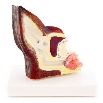
- Individual organ models: Hearts, eyes, ears, skin, digestive organs, and more—some featuring pathological details such as parasites or infections.
- Modular and dual-sided designs: Many models are detachable or reversible, allowing a detailed look at healthy vs. affected anatomical states.

Detailed Features for Veterinary Education
Many of our models are designed with dual views—external structure on one side, internal cross-section on the other. Ear models may show normal anatomy on one side and diseased tissue on the reverse. Fully disassemblable models allow for advanced study of organ placement, relationships, and system-level function. These tools make veterinary consultations and academic instruction far more effective and engaging.
Order veterinary animal and internal organ models from Veteq Group for fast UK delivery and expert support. Each model is selected for realism, educational value, and professional-grade durability.