


|
Quantity
|
Out of stock
|
||
|
|
|||
|
|
|||
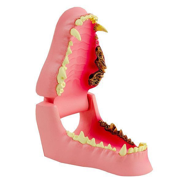
Large Canine Jaw Anatomical Model
Educational Dental Model Showing Healthy and Diseased Dentition
The Large Canine Jaw Model offers a detailed anatomical comparison between healthy and pathological dental conditions in dogs. Designed for veterinary professionals, educators, and clinical consultations, this model accurately represents a range of common oral health issues on the left side, while displaying healthy dentition on the right.
Two-Sided Design: Healthy vs. Pathological Dentition
The model can be viewed both open and closed, making it a practical tool for explaining canine oral anatomy and dental disorders to pet owners and students. The contrasting sides help reinforce the importance of regular dental care in dogs.
Conditions Represented on the Diseased Side:
- Broken tooth
- Periodontitis
- Gum inflammation
- Gingival recession
- Plaque
- Tartar (calculus)
- Worn incisors
- Missing premolar
- Persisting deciduous teeth

Key Features
- Right side: healthy canine dentition
- Left side: dental pathologies for comparison
- Openable and closable jaw model for interactive demonstration
- Ideal for clinical explanation and educational training
Specifications
- Dimensions (H × W × D): approx. 12.1 × 7 × 5.1 cm
Applications
- Veterinary dentistry education and consultation
- Client education on canine dental hygiene and oral health
- Use in veterinary universities and dental training courses
Why Choose This Model?
The Large Canine Jaw Anatomical Model is a practical and effective tool for visualizing dental health vs. disease. With lifelike detail and a functional open/close design, it supports improved communication between veterinarians and pet owners and enhances dental education programs.